⚕ Clinician & Trainee Reference ASN POCUS · KDIGO Aligned Bedside · Four Domains

Point-of-Care Ultrasound
in Nephrology

A bedside field guide — applied pathophysiology with a probe: image → mechanism → cross-organ consequence → action.

PublishedNailathalaGipatikPepalwal: ReferencesMga SanggunianMga TinubdanReng Reperensya: 11 Read timeOras ng pagbasaOras sa pagbasaOras ning pamamasa: Audience: Nephrology fellows · IM residents · practicing nephrologists & hospitalists
Circular vignette hero illustration for the POCUS in nephrology field guide — a clinician's hand holding a curvilinear ultrasound probe over a translucent kidney with overlaid B-lines and a Doppler waveform, in calm clinical light.
Bedside use only. POCUS is a focused, hypothesis-driven exam — it confirms or refutes one physiologic question and translates the answer into a therapeutic decision. It is not a substitute for formal radiology ultrasound or a complete echocardiogram, and competence requires supervised acquisition logs (§7). The clinical thresholds here (VEXUS, IVC/RAP, rule-of-6s, PVR, RI) reflect current published consensus; verify against your institution's protocols before use.

Best used when — at a glance

Physics, Probes, Knobology & Artifact Literacy

The minimum bench you need before approaching any window. Every higher-yield interpretation downstream is enabled — or sabotaged — by these basics.

POCUS in nephrology is built on three habits: match the probe to the question, optimize the image before interpreting it, and read artifact as information, not noise. Skipping any of these turns a confident bedside read into a confidently wrong one.1,3

1.1 Physics in plain terms

Ultrasound is the trade between frequency (higher = better axial resolution, worse penetration) and depth. A 2–5 MHz curvilinear beam reaches the deep retroperitoneum and IVC; a 7–12 MHz linear beam resolves a 2 mm fistula vessel wall but stops in the subcutaneous tissue. Axial resolution is the smallest distance the probe can separate along the beam (limited by wavelength); lateral resolution is across the beam (limited by beam width — sharper at the focal zone). Attenuation is the loss of signal with depth; gain (and time-gain compensation, TGC) is how we restore it without amplifying noise.

1.2 Probe selection — three transducers, three jobs

ProbeFrequencyNephrology use
Curvilinear2–5 MHzKidney, bladder, IVC long-axis, hepatic vein, intrarenal Doppler — the workhorse of renal & volume POCUS.
Phased array1–5 MHzFocused cardiac (PLAX/PSAX/A4C/subcostal) and subcostal IVC through a small footprint between ribs.
Linear (high-frequency)7–12 MHzAV fistula/graft surveillance, pre-creation vascular mapping, central venous access — superficial, high-resolution.

1.3 Knobology — the five dials that matter at the bedside

  • Depth — set so the structure of interest fills two-thirds of the screen.
  • Gain & TGC — gain raises the whole image; TGC tunes near vs far. Aim for a fluid-filled vessel that is truly anechoic, not "dirty black."
  • Focal zone — place at the depth of the target (sharpest lateral resolution there).
  • Presets — abdominal for kidney/IVC; cardiac for focused echo (reverses default orientation marker); vascular for fistula.
  • Doppler — color box small and tilted ≤ 60° to flow; pulsed-wave gate inside the vessel; lower PRF for slow venous signals, higher for arterial.
Figure 1 — POCUS probe selection. Three transducers (curvilinear 2–5 MHz, phased array 1–5 MHz, linear 7–12 MHz) mapped to their nephrology applications with grayscale B-mode thumbnails per use case, plus a frequency-vs-penetration curve showing kidney/IVC, focused-echo, and AV-access target depths.
Figure 1. Probe selection by depth and indication — curvilinear (kidney, IVC, bladder), phased array (focused echo, subcostal IVC), linear (AV access, IJ, vascular mapping). Pick the lowest frequency that resolves your target.

Doppler pearl — intrarenal resistive index (RI)

RI = (PSV − EDV) / PSV; normal intrarenal RI < 0.70. RI is non-specific but supports parenchymal/vascular disease and rises with venous congestion — pair it with the VEXUS module (§3) before acting on it in isolation.

1.4 Artifact literacy — read what isn't really there

  • Acoustic shadowing — hyperechoic surface (stone, calcification, rib) + black "void" deep to it. Diagnostic for a calculus or rib edge.
  • Posterior enhancement — bright signal deep to a fluid-filled structure (cyst, full bladder). Distinguishes simple cyst from solid mass.
  • Reverberation — repeating horizontal lines below a strong reflector. In the lung this is the normal A-line.
  • Ring-down (B-line) — vertical laser-like line from the pleural line to the screen's bottom edge, erasing A-lines, moving with sliding. Marker of extravascular lung water (§4).
  • Mirror artifact — a duplicate of liver/spleen across the diaphragm — a common pitfall when judging pleural effusion.

1.5 Governance — image archiving, infection control, scope

Every bedside study should be archivable: a representative still or short clip per window, labeled with patient identifier and indication. Probes are semi-critical when they contact intact mucosa (e.g. transvaginal — not used here) and non-critical for intact skin: low-level disinfectant wipe (manufacturer-approved) between every patient; high-level disinfection for sterile-field use (e.g. internal jugular cannulation with sterile sheath). Scope of practice is institutional — POCUS findings should be documented as focused (not "complete US"), and any uncertain finding triggers formal imaging.2

Obstruction, Parenchyma & the Bladder

The single highest-yield application for the new clinician on the AKI ward: rule in obstruction, read chronicity off the cortex, and reframe "renal failure" as retention when the bladder is the problem.

2.1 The four bedside questions

  • Is there obstruction? (hydronephrosis — and is the bladder full?)
  • Is this acute or chronic kidney disease? (size, cortex thickness, echogenicity)
  • Is the bladder the problem? (post-void residual)
  • Is a mass cystic or solid? (simple vs complex; when to escalate)

2.2 Acquisition

  • Right kidney via the hepatic acoustic window: probe at the mid-axillary line, coronal orientation, marker cephalad. Use the liver as the sonographic "lens." Sweep through the kidney in long axis, then rotate 90° for short axis.
  • Left kidney via the splenic window: more posterior and cephalad than the right (the spleen is smaller; bowel gas often obscures). Have the patient take a deep breath; roll into right lateral decubitus if needed.
  • Bladder: suprapubic, transverse first to find the maximal AP and transverse dimensions, then sagittal for craniocaudal. Volume ≈ 0.52 × W × H × D (mL).
Figure 2 — Hydronephrosis POCUS grading. Three side-by-side grayscale B-mode panels (mild, moderate, severe) with paired coronal-anatomy insets, showing progression from a dilated pelvis with preserved cortex (mild) → rounded bear-paw calyces with early thinning (moderate) → ballooned anechoic calyces with marked cortical thinning (severe). Each panel carries a cortical-thickness caliper.
Figure 2. Hydronephrosis grades (mild · moderate · severe). Grade reflects both degree and chronicity of obstruction — cortical thinning marks chronicity. Relieve before pursuing intrinsic AKI workup.

2.3 Interpretation & thresholds

FindingTeaching point / threshold
Hydronephrosis gradingMild — pelvis & calyces dilated, cortex preserved.
Moderate — rounded ("bear-paw") calyces, mild cortical thinning.
Severe — ballooned calyces, marked cortical thinning. Grade reflects both degree and chronicity of obstruction.
Kidney sizeNormal ~9–12 cm bipolar length. Small (< 9 cm), echogenic, thin cortex → chronicity (CKD). Normal or enlarged kidneys with AKI suggest an acute, potentially reversible process.
EchogenicityCortex is normally hypo-echoic to adjacent liver/spleen. Cortex ≥ liver echogenicity → medical renal disease (glomerular/tubulointerstitial).
CystSimple — anechoic, thin wall, posterior enhancement → benign. Complex (septa, mural nodule, solid component, calcification) → formal imaging / Bosniak classification.
Bladder / PVRPost-void residual normal < 50 mL (< 65 y) to < 100 mL (≥ 65 y). A large PVR reframes "renal failure" as post-renal/retention — catheter first, work-up second.
StonesHyperechoic focus + posterior acoustic shadow. Color Doppler twinkle artifact supports a calculus when shadow is equivocal.

Decision point — AKI workup

An obstructed (hydronephrotic) kidney or a high PVR converts the differential to post-renal AKIdecompress first (Foley / nephrostomy) before pursuing intrinsic causes. Absence of hydronephrosis does not fully exclude early or volume-depleted obstruction; rescan after rehydration or pursue CT urography if pre-test suspicion stays high.

Figure 3 — Bladder volume and post-void residual measurement. Suprapubic transverse B-mode panel (Width × Transverse calipers) + sagittal B-mode panel (Height caliper) + formula card showing Volume ≈ 0.52 × W × T × H (mL) with PVR thresholds and the retention decision point.
Figure 3. Bladder volume from three orthogonal diameters. PVR < 50 mL (< 65 y) or < 100 mL (≥ 65 y) is normal; higher values reframe "renal failure" as retention — catheterize first.

2.4 Pitfalls & mimics

  • Extrarenal pelvis — a pelvis that lies outside the renal sinus is normally a few mL of fluid and mimics mild hydronephrosis. Reassess after voiding.
  • Parapelvic cysts — clustered, round, do not communicate with the ureter; look for the cyst wall vs branched calyceal anatomy.
  • Full bladder / reflux — a distended bladder back-pressures the upper tract and falsely "creates" mild bilateral dilation. Rescan after catheter drainage.
  • Decompressed obstruction — early or volume-depleted obstruction may show minimal dilation; bowel gas commonly limits the left kidney.
  • Renal cell carcinoma — a solid renal lesion is never a POCUS diagnosis; refer for contrast-enhanced CT/MRI.

Cross-organ link — post-renal AKI

Obstruction raises intratubular pressure → transcapillary filtration gradient collapses → GFR falls. Relief restores filtration over hours to days. When obstruction co-exists with venous congestion (high VEXUS, §3), both axes must be relieved for renal recovery — neither alone is enough.

The Highest-Yield Nephrology POCUS Domain

Replace "dry weight" guesswork with a graded, mechanism-based read of forward filling and backward venous congestion. This is the sonographic face of cardiorenal syndrome.

3.1 IVC — right atrial pressure estimate

The IVC is a low-pressure venous capacitance vessel that mirrors right atrial pressure (RAP) through its diameter and respiratory variation. Image the IVC 2–3 cm caudal to the cavo-atrial junction (or just caudal to the IVC–hepatic vein confluence) in long axis, with the probe in the subxiphoid window. Measure diameter at end-expiration and assess collapse with a sharp sniff.

IVC patternInterpretation (RAP estimate)
≤ 2.1 cm, > 50% collapse with sniffNormal RAP (~3 mmHg, range 0–5)
Intermediate (either ≤ 2.1 cm with poor collapse, or > 2.1 cm with brisk collapse)Intermediate RAP (~8 mmHg) — integrate with other windows
> 2.1 cm, < 50% collapseElevated RAP (~15 mmHg) — plethoric IVC

Confounders. Mechanical ventilation (positive pleural pressure inverts the normal respiratory variation), raised intra-abdominal pressure (transmits to IVC), pulmonary hypertension, athletic physiology, and severe tricuspid regurgitation all distort the diameter–pressure relationship. Read the IVC as a gate, not the whole story.4

Figure 4 — IVC long-axis ultrasound panel with a caliper 2–3 cm caudal to the cavo-atrial junction, paired with a three-tier RAP estimate table (≤ 2.1 cm with > 50% collapse → ~3 mmHg; intermediate → ~8 mmHg; > 2.1 cm with < 50% collapse → ~15 mmHg) and a confounder list (mechanical ventilation, intra-abdominal pressure, pulmonary hypertension, athletes, tricuspid regurgitation).
Figure 4. IVC diameter + respiratory variation → RAP tier. Measure 2–3 cm caudal to the hepatic-vein confluence; integrate with other windows when confounders apply.

3.2 VEXUS — Venous Excess Ultrasound grading

VEXUS converts IVC plethora into a graded venous-congestion score by adding pulsed-wave Doppler of the hepatic vein, portal vein, and intrarenal vein. It tracks dynamically across hemodialysis ultrafiltration (ACUVEX cohort)5 and predicts worsening renal function in acute heart failure6 and the ICU AKI prevalence cohort.7

Component Doppler severity

VeinNormalMild abnormalitySevere abnormality
HepaticS > D (systolic dominant, both antegrade)S < D (S still antegrade)S-wave reversal (retrograde flow toward liver)
PortalPulsatility < 30%30–49%≥ 50% pulsatility
Intrarenal veinContinuousBiphasic (S + D waves)Monophasic (D-only)8

Grade assignment

GradeDefinition
Grade 0IVC < 2 cm — no congestion; do not grade.
Grade 1 — MildIVC ≥ 2 cm plus normal or only mild Doppler abnormalities.
Grade 2 — ModerateIVC ≥ 2 cm plus severe abnormality in exactly one venous bed.
Grade 3 — SevereIVC ≥ 2 cm plus severe abnormality in ≥ 2 venous beds.
Figure 5 — VEXUS four-grade Doppler ladder. Four rows (Grade 0 → 3, top-to-bottom, color-coded green → amber → orange → red) with three columns of pulsed-wave Doppler waveforms (hepatic, portal, intrarenal) and an IVC reference at left. Progression: Grade 0 IVC < 2 cm, hepatic S > D, portal pulsatility < 30%, renal continuous → Grade 3 IVC ≥ 2 cm with hepatic S-wave reversal, portal ≥ 50% pulsatility, monophasic intrarenal.
Figure 5. VEXUS — Venous Excess Ultrasound grading. Plethoric IVC + severe abnormality in ≥ 2 venous beds = Grade 3. Higher grade ⇒ backward venous congestion ⇒ falling GFR ⇒ decongest and re-scan.

Pitfalls — when not to trust the grade

  • Cirrhosis / high-output states produce portal pulsatility unrelated to right-heart congestion.
  • Arrhythmia (AF, frequent ectopy) degrades all three waveforms — interpret with caution.
  • Renal vein signal is technically demanding; use a small color box at an interlobar vein, sample with a 60° angle and a low velocity scale.
  • Require IVC plethora (≥ 2 cm) before grading — Doppler abnormalities without a plethoric IVC are not VEXUS.

Decision point — congestive nephropathy

A high VEXUS grade signals that renal dysfunction is driven by backward venous congestion (elevated renal venous pressure → reduced transrenal perfusion gradient → falling GFR), not "pre-renal underfilling." The action is decongestion — loop diuretics, ultrafiltration, or both — and a falling VEXUS grade is an objective endpoint. Avoid the reflex fluid bolus.

3.3 Cross-organ link

VEXUS is the sonographic face of cardiorenal syndrome (CRS) types 1 & 2, and of hepatic congestion in the same patient. The unifying frame: the kidney is an early victim of systemic venous congestion. When VEXUS is paired with lung ultrasound (§4) and a focused echo, the volume status of the AHF or HD patient is mapped end-to-end at the bedside.

Extravascular Lung Water & the Four-Window Focused Echo

Lung-ultrasound B-lines see pulmonary congestion before auscultation, radiograph, or symptoms. The focused echo answers four binary questions that change therapy at the bedside.

4.1 Lung ultrasound — A-lines vs B-lines

Set the curvilinear (or phased) probe perpendicular to the chest wall in a longitudinal orientation so two rib shadows flank a bright pleural line ("bat sign"). The lung sits deep to the pleura.

  • A-lines — horizontal lines parallel to and equidistant below the pleural line. Reverberation artifact = normal aerated lung or pneumothorax (if sliding is also absent).
  • B-lines — vertical, hyperechoic, laser-like lines arising from the pleural line, extending to the bottom of the screen, erasing A-lines, moving with sliding. They mark interstitial fluid (extravascular lung water, EVLW).
  • Quantification. ≥ 3 B-lines in a single intercostal field = positive zone. Sum positive zones across an 8-zone (4 per hemithorax) or 28-zone protocol for a semi-quantitative congestion score.
Figure 6 — Lung POCUS. Left half: two grayscale B-mode panels side-by-side showing A-lines (horizontal reverberation, dry aerated lung) vs B-lines (≥ 3 vertical laser-like ring-down lines from the pleura to the screen edge, interstitial fluid). Right half: anterior chest torso with an 8-zone grid (4 zones per hemithorax) plus a small 28-zone reference inset. Bat sign visible on each US panel.
Figure 6. A-lines vs B-lines + zone scoring for extravascular lung water. ≥ 3 B-lines in a single intercostal field = positive zone; sum across 8 or 28 zones for the congestion score.

Why this matters in nephrology

B-lines detect pulmonary congestion before rales, before chest X-ray opacities, and before dyspnea. LUS-guided ultrafiltration in HD is associated with better BP control and fewer decompensation events, and a high B-line burden flags an undertreated cardiorenal phenotype that fluid restriction alone will not fix.9

Also screen for

  • Pleural effusion — anechoic stripe above the diaphragm with the "spine sign" (vertebrae visible above the diaphragm because of the fluid acoustic window).
  • Pneumothorax — absent pleural sliding + absent B-lines + a "lung point" where sliding resumes. (Uncommon in pure nephrology workflow but important after access / biopsy.)

4.2 Focused cardiac POCUS

Four windows, four questions — none of them an echocardiographer's exam.

WindowTargetBedside question
PLAX (parasternal long-axis)LV, mitral & aortic valves, LA, pericardiumGross LV systolic function (eyeball EF); pericardial effusion.
PSAX (parasternal short-axis, mid-papillary)LV cavity geometry & thickeningRegional wall motion at a glance; "D-shaped" septum → RV pressure/volume overload.
A4C (apical 4-chamber)All four chambers; tricuspid & mitral inflowRV size relative to LV (RV ≥ LV at the base = abnormal); gross atrial size.
Subcostal4-chamber + IVC long-axisRescue view when PLAX/A4C fail (COPD, post-op); IVC for RAP (§3).

Pericardial effusion / tamponade physiology

Anechoic stripe surrounding the heart inside the bright pericardium. Tamponade is a clinical diagnosis, but POCUS supports it: a circumferential, often large effusion, with right-atrial systolic collapse, right-ventricular early-diastolic collapse, and a plethoric, non-collapsing IVC. In a uremic patient with hypotension, this is a stat-call sign.

Figure 7 — Focused cardiac POCUS, four windows in a 2×2 grid. PLAX (LV, MV/AV, pericardium), PSAX mid-papillary (LV cavity geometry, D-shaped septum), A4C (all four chambers, RV:LV ratio), and subcostal (rescue view + IVC). Each panel carries a small torso inset showing probe placement and orientation marker.
Figure 7. Four focused-echo windows, one question each: gross LV function (PLAX), septal geometry / RV overload (PSAX), RV:LV ratio (A4C), rescue + IVC (subcostal).

Decision point — focused echo limits

Focused cardiac POCUS rules in gross findings; it does not exclude regional wall motion abnormality, valvular disease, or HFpEF. When the bedside question is "is the kidney being injured by a failing heart?", a normal focused echo does not close the workup — refer for full echocardiography.

AV Access Surveillance & Ultrasound-Guided Procedures

The dialysis patient's lifeline is sonographic. Early access surveillance preserves it; procedural guidance prevents catheter-related morbidity.

5.1 AV access surveillance (linear probe)

Use the high-frequency linear probe across the fistula/graft in short axis first (round, anechoic lumen with thin echogenic wall) to locate the anastomosis, then long axis for length and flow direction. Add color Doppler (low PRF for venous outflow, high PRF for arterial inflow) and PW Doppler for peak velocities.

Rule of 6s — AV fistula maturation

  • Flow > 600 mL/min in the draining vein.
  • Diameter ≥ 6 mm at the cannulation segment.
  • Depth ≤ 6 mm from skin (deeper = cannulation difficulty).
  • Assess at ~6 weeks post-creation.
  • Venous needle points away from the anastomosis; left arm preferred when possible.

What to detect

  • Juxta-anastomotic / outflow stenosis — > 50% diameter reduction or a focal > 2:1 peak-velocity ratio across the narrowing; visible aliasing on color Doppler.
  • Thrombosis — non-compressible lumen, no color or PW signal, possibly echogenic intraluminal material.
  • Aneurysm / pseudoaneurysm — focal saccular dilation; yin-yang color flow inside a pseudoaneurysm with a feeding "neck."
  • Steal — high-volume access with distal arterial flow reversal, particularly in diabetic / proximal accesses (clinical + perfusion findings still primary).
  • Pre-creation vascular mapping — arterial inflow (radial / brachial) calibre & patency; venous outflow (cephalic / basilic) calibre, patency, and depth.

5.2 Procedural guidance

  • Central venous accessreal-time ultrasound-guided cannulation of the internal jugular and femoral veins is now standard of care: pre-scan for patency, thrombus, and a compressible vein, then keep the needle tip in view through the wall. (Subclavian is anatomic, not US-guided.)
  • Kidney biopsy — patient prone, breath-held expiration; target the lower-pole cortex; confirm no overlying bowel loop, large vessel, or pleural reflection. Real-time guidance (out-of-plane or in-plane) is now preferred to a single pre-scan "mark and stab."
  • PD catheter siting — ultrasound-marked entry to avoid epigastric vessels and adhesions; intra-procedural check for the catheter tip in the pelvis.
  • Difficult bladder catheterization — sonographic confirmation that the bladder is actually full before another pass; rules out the empty-bladder false-failure.
Figure 8 — Mature left-arm radial-cephalic AV fistula schematic with rule-of-6s callouts (flow > 600 mL/min, diameter ≥ 6 mm, depth ≤ 6 mm, assess at ~6 weeks) and three complication mini-panels on the right: juxta-anastomotic stenosis (> 2:1 PSV ratio, color aliasing), thrombosis (non-compressible, no flow), and pseudoaneurysm (yin-yang sign with feeding neck).
Figure 8. AV fistula rule of 6s + complication panels. Venous needle points away from the anastomosis; left arm preferred when possible.

Cross-organ link — access is the dialysis lifeline

Early sonographic surveillance — a brief monthly look at flow, depth, and any focal velocity step-up — preserves AV access patency and pre-empts thrombosis. Every catheter avoided is a downstream CRBSI, central-vein stenosis, and hospitalization avoided.

The Five-Point Scan → Hemodynamic Phenotype → Action

The capstone module. Findings from each window are assembled into one of a few physiologic phenotypes, each with a guideline-aligned action. This is what protects the patient from "fluid in both directions."

Figure 9 — Integrated POCUS algorithm. Top row: five-point scan strip (kidney/bladder · IVC · lungs · cardiac · VEXUS). Center: five phenotype pathways with POCUS signatures and guideline-anchored actions — congestive nephropathy/CRS (decongest), cardiorenal with pump failure (decongest + neurohormonal), true hypovolemia (volume resuscitate), obstructive/post-renal AKI (decompress first), and tamponade physiology (stat pericardiocentesis).
Figure 9. Five-point scan → hemodynamic phenotype → guideline-anchored action. The kidney lives between forward perfusion and backward congestion; POCUS lets you see both sides at the bedside.

6.1 The five-point scan

In any undifferentiated AKI, suspected congestion, or pre-dialysis assessment, scan five sites in this order and record one finding per site:

  1. Kidneys / bladder — hydronephrosis? PVR?
  2. IVC — diameter and collapse → RAP tier.
  3. Lungs — 8-zone B-line count.
  4. Focused cardiac — LV "eyeball" EF, pericardial effusion, RV size.
  5. VEXUS Doppler — hepatic, portal, intrarenal vein (only if IVC ≥ 2 cm).

6.2 Phenotype → action

PhenotypePOCUS signatureAction (guideline anchor)
Congestive nephropathy / CRS-1/2Plethoric IVC, VEXUS 2–3, ≥ 3 B-lines per zone in multiple zones, ± reduced LV functionDecongest — loop diuretic ± ultrafiltration; reassess by falling VEXUS / B-line burden. SGLT2 inhibitor where indicated (KDIGO 2024 / ADA / ACC).
True hypovolemia / pre-renalSmall, collapsible IVC; dry lungs (A-pattern); hyperdynamic LV ("kissing walls")Volume resuscitation (balanced crystalloid), reassess dynamically; stop pressors that mask the deficit.
Obstructive / post-renal AKIHydronephrosis ± high PVR (full bladder)Decompress first (Foley / nephrostomy) — then revisit the intrinsic workup.
Cardiorenal with pump failureReduced LV, B-lines, high VEXUS, possibly pericardial effusionDecongest + neurohormonal therapy (ARNI/MRA/SGLT2i per ACC/AHA HF); avoid the reflex fluid bolus.
Tamponade physiologyPericardial effusion + RA/RV diastolic collapse + plethoric IVCStat pericardiocentesis — fluid is a temporizing bridge, not a treatment.

Unifying frame — the kidney between forward perfusion and backward congestion

POCUS lets the clinician see both sides of the kidney's perfusion equation at the bedside and titrate therapy to an objective, repeatable endpoint — not to weight, symptoms, or a static creatinine alone. This is the single biggest cognitive shift POCUS brings to nephrology: volume is a phenotype, not a number on a scale.

6.3 An integrated example

A 68-year-old with HFrEF and CKD-G3b is admitted with worsening creatinine. Reflex instinct: "pre-renal — give fluids." Five-point scan: no hydronephrosis, IVC 2.6 cm with < 20% collapse, 4 B-lines per zone bilaterally in lower zones, mildly reduced LV with a moderately dilated RV, VEXUS Grade 3 (S-reversal in hepatic, portal pulsatility 60%, monophasic intrarenal). The action changes entirely: decongestion, not volume; SGLT2i continued; falling VEXUS at 48 h becomes the objective endpoint.

Tiered Skills, Logbooks & Departmental QA

POCUS is a skill, not a credential — but it must be governed. A reproducible competency pathway protects patients and trainees alike.

Documented training gap: in published surveys, only ~38% of nephrology fellows received POCUS education and only ~33% of those felt competent to perform it independently.10 This guide is a self-study scaffold; it does not replace supervised acquisition.

7.1 Tiered competency

  1. Tier 1 — Image acquisition. Can the trainee obtain a diagnostic-quality image at each window? Direct-observation checklist per application.
  2. Tier 2 — Interpretation. Can the trainee distinguish normal from abnormal and label findings against published thresholds (e.g. VEXUS grades, RAP tiers, rule of 6s)?
  3. Tier 3 — Clinical integration. Can the trainee combine multi-organ findings into a phenotype and a guideline-aligned action — and articulate what would change it?

7.2 Image logbook

  • Suggested minimum supervised studies per domain before independent use (institutional thresholds vary; ASN precourse offers a reasonable target).2
  • Archive representative still images and short clips (with patient identifier, indication, and a one-line interpretation) for every supervised case.
  • Quarterly portfolio review with a designated POCUS faculty mentor.

7.3 Assessment

  • Entrustable tasks — directly observed acquisition + interpretation.
  • Image-review quizzes — graded with annotated answer keys.
  • Integrated case OSCEs — the trainee performs a five-point scan and proposes a phenotype + action.

7.4 Governance — a departmental POCUS committee

  • Owns credentialing criteria and the privileging pathway.
  • Runs QA image review (random audits of archived studies).
  • Maintains the curriculum and the institutional protocol pack (probe disinfection, archiving, reporting language).
  • Mirrors published ASN recommendations for nephrology POCUS programs.2

Limits and when to escalate

A POCUS finding that you cannot reconcile with the clinical picture — or any solid renal lesion, complex cyst, unexpected pleural fluid, suspected aortic dissection / abdominal aortic aneurysm, or any pediatric uncertainty — is a referral for formal radiology imaging or echocardiography. POCUS is the bridge to the question; it is not always the answer.

Visual Atlas — Probes, Grades, Doppler & Phenotypes

Nine planned figures map the curriculum visually. Each is a Constitution-v1.0 hybrid: anatomical truth + ultrasound physics + a clinical-action callout, dual clinician/patient labels, navy williamriveromd.com watermark.

8.1 Quick-reference card

DomainThreshold / rule
HydronephrosisMild = pelvis/calyces dilated, cortex preserved · Moderate = rounded calyces, mild cortical thinning · Severe = ballooned calyces, marked thinning
Kidney sizeNormal ~9–12 cm · Small + echogenic → CKD · Normal/enlarged in AKI → potentially reversible
PVRNormal < 50 mL (< 65 y); < 100 mL (≥ 65 y)
IVC → RAP≤ 2.1 cm + > 50% collapse = ~3 mmHg · > 2.1 cm + < 50% = ~15 mmHg · intermediate = ~8 mmHg
VEXUSGrade 0 (IVC < 2 cm) · 1 mild · 2 severe in one bed · 3 severe in ≥ 2 beds
Lung B-lines≥ 3 per intercostal field = positive zone; sum 8 or 28 zones for score
AV fistula rule of 6sFlow > 600 mL/min · diameter ≥ 6 mm · depth ≤ 6 mm · at ~6 weeks
Intrarenal RINormal < 0.70; elevated supports parenchymal/vascular disease & congestion

8.2 Figure manifest

#FilenameSubject
F1pocus-probe-selection-visual-aid-hybrid-v2.pngCurvilinear / phased-array / linear probes matched to kidney, IVC/cardiac, AV access; frequency-vs-penetration trade-off.
F2 ★hydronephrosis-pocus-grading-hybrid-v2.pngMild / moderate / severe hydronephrosis with paired anatomy & US physics (FLAGSHIP).
F3bladder-pvr-measurement-visual-aid-hybrid-v2.pngSuprapubic bladder transverse + sagittal; three-dimension volume formula; PVR thresholds.
F4ivc-rap-assessment-hybrid-v2.pngIVC long-axis with caliper, collapsibility, three-tier RAP table, confounder list.
F5 ★vexus-grading-hybrid-v2.pngVEXUS four-grade Doppler ladder — hepatic, portal, intrarenal (FLAGSHIP).
F6 ★lung-blines-evlw-hybrid-v2.pngA-lines vs B-lines, bat sign, 8-zone & 28-zone grids (FLAGSHIP).
F7focused-cardiac-windows-visual-aid-hybrid-v2.pngPLAX / PSAX / A4C / subcostal with target per window.
F8av-fistula-rule-of-6s-hybrid-v2.pngFistula anatomy with rule-of-6s callouts; stenosis / thrombosis / aneurysm panels.
F9pocus-volume-phenotypes-visual-aid-hybrid-v2.pngFive-point scan flow → phenotype → action decision tree.

Figure system note

Each figure follows the williamriveromd.com Constitution v1.0 (1672×941 px, 16:9, GPT-4o), with the mandatory seven-layer architecture (anatomical truth · spatial orientation · render specifications · imaging physics · negative constraints · style reference · QA checklist). Full prompts for the three flagship figures (F2, F5, F6) are maintained in the blueprint and are paste-ready into the image generator.

Glossary of Terms & Abbreviations

Every acronym and sonographic term used in this guide, with a one-line working definition. Skim before rounds; refer back during a scan.

9.1 Acronyms — physiology, syndromes & programs

TermExpansionWorking definition
POCUSPoint-of-Care UltrasoundFocused, hypothesis-driven bedside US — confirms or refutes one physiologic question and converts the answer into a therapeutic decision.
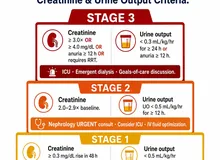
AKIAcute Kidney InjuryAbrupt fall in GFR over hours–days, staged by KDIGO creatinine/urine-output criteria.
CKDChronic Kidney DiseaseGFR < 60 mL/min/1.73 m² or marker of kidney damage ≥ 3 months.
GFRGlomerular Filtration RateVolume of plasma filtered per unit time; estimated (eGFR) by CKD-EPI or similar.
AHFAcute Heart FailureRapid onset/worsening of HF signs/symptoms requiring urgent therapy.
HF / HFrEF / HFpEFHeart Failure / reduced / preserved EFReduced (LVEF ≤ 40%) vs preserved (LVEF ≥ 50%) ejection fraction.
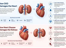
CRS-1 / CRS-2Cardiorenal Syndrome types 1, 2Acute (1) or chronic (2) cardiac dysfunction driving renal dysfunction — VEXUS is its sonographic face.
LVHLeft Ventricular HypertrophyWall thickening of the LV, often driven by hypertension or volume overload.
LV / RV / LA / RALeft / Right Ventricle, AtriumThe four cardiac chambers as labeled on every echo window.
EFEjection FractionStroke volume ÷ end-diastolic volume — "eyeball" on POCUS, quantified on formal echo.
RAPRight Atrial PressurePressure in the RA; estimated from IVC diameter + respiratory variation (§3).
TRTricuspid RegurgitationBackward flow across the tricuspid valve — a confounder of IVC interpretation.
HDHemodialysisExtracorporeal renal-replacement therapy by diffusion ± convection.
PDPeritoneal DialysisRRT using the peritoneum as the dialysis membrane.
IDHIntradialytic HypotensionBP drop during HD; mechanism-mapped to refilling, tonicity, tone, or cardiac reserve.
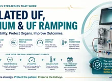
UF / UFRUltrafiltration / Ultrafiltration RateConvective fluid removal during HD; UFR usually expressed in mL/kg/hr.
IDWGInterdialytic Weight GainWeight gained between HD sessions, mostly fluid.
CRBSICatheter-Related Bloodstream InfectionBacteremia attributable to an intravascular catheter.
IJInternal Jugular (vein)The preferred US-guided central venous access site.
AVF / AVGArteriovenous Fistula / GraftSurgical (AVF) or prosthetic (AVG) anastomosis used for HD access.
KDIGOKidney Disease: Improving Global OutcomesInternational nephrology guideline body (AKI, CKD, BP, AKI/AKD, diabetes, etc.).
ACC / AHAAmerican College of Cardiology / Heart AssociationU.S. cardiology guideline bodies (HF, AHF, pericardial disease).
ADAAmerican Diabetes AssociationDiabetes guideline body — SGLT2i indications in CKD/HF.
ASNAmerican Society of NephrologyKidney Week and the POCUS precourse cited in §7.
SGLT2iSodium-Glucose Cotransporter-2 inhibitorClass with renal & cardiac protection (dapa-, empa-, cana-gliflozin).
ARNIAngiotensin Receptor–Neprilysin InhibitorSacubitril/valsartan — pillar of HFrEF therapy.
MRAMineralocorticoid Receptor AntagonistSpironolactone / eplerenone / finerenone — HF + CKD/diabetic kidney disease.
OSCEObjective Structured Clinical ExaminationPerformance-based assessment used in §7 to test integrated POCUS skills.
QAQuality AssuranceDepartmental image review & credentialing process for POCUS programs.

9.2 VEXUS, IVC & venous Doppler vocabulary

TermDefinition
VEXUSVenous Excess Ultrasound — a four-grade venous-congestion score combining IVC plethora with hepatic, portal, and intrarenal vein pulsed-wave Doppler.
IVCInferior Vena Cava — measured 2–3 cm caudal to the cavo-atrial junction in long axis.
Plethoric IVCIVC ≥ 2 cm with < 50% respiratory collapse — prerequisite to grade VEXUS.
S / D / A wavesHepatic-vein Doppler components: systolic (S, antegrade in atrial diastole), diastolic (D, antegrade in atrial systole), and the A reversal in late atrial systole.
S-wave reversalHepatic-vein S wave flows retrograde toward the liver — severe right-heart congestion (VEXUS Grade 3 hepatic).
Portal pulsatility (%)(Vmax − Vmin) ÷ Vmax × 100 in the main portal vein. < 30% normal; 30–49% mild; ≥ 50% severe.
Continuous / biphasic / monophasic intrarenal veinNormal / mild / severe venous congestion at the interlobar level; monophasic-D-only is the most ominous.
RIResistive Index = (PSV − EDV) / PSV; normal intrarenal < 0.70.
PSV / EDVPeak Systolic / End-Diastolic Velocity on PW Doppler — the inputs to RI.
PW / CW DopplerPulsed-wave (range-gated, used for vessel sampling) vs continuous-wave (high velocities, no range gating).
Color DopplerVelocity-encoded color overlay; aliases above the Nyquist limit (set by PRF).
PRFPulse Repetition Frequency — set low for slow venous flow, high for arterial.
AliasingColor "wrap-around" or PW wraparound when flow velocity exceeds the Nyquist limit (PRF / 2).
Insonation angleThe angle between the Doppler beam and flow; keep ≤ 60° for accurate velocity quantification.

9.3 Renal, bladder & obstruction terms

TermDefinition
HydronephrosisDilation of the renal pelvis and calyces — sonographic surrogate for downstream obstruction.
Bear-paw signRounded, interconnected calyces in moderate hydronephrosis.
Extrarenal pelvisRenal pelvis lying outside the sinus — normal variant; mimics mild hydronephrosis.
Parapelvic cystSimple cyst clustered near the renal sinus; can mimic dilated calyces (but doesn't communicate).
Cortex / medulla / sinusOuter parenchyma (cortex), inner pyramids (medulla), central echogenic fat/vessels/collecting system (sinus).
Cortical echogenicityBrightness of cortex relative to liver/spleen — cortex normally hypo-echoic; ≥ liver suggests medical renal disease.
Twinkle artifactRandom color signal behind a calcification on color Doppler — supports a calculus when shadowing is equivocal.
PVRPost-Void Residual urine volume; normal < 50 mL (< 65 y) to < 100 mL (≥ 65 y).
Bosniak classificationCT/MRI-based risk stratification for complex renal cysts (I → IV); POCUS does not grade Bosniak.
Post-obstructive diuresisLarge urine output after relief of obstruction; requires careful electrolyte and volume management.

9.4 Sonographic physics & artifact terms

TermDefinition
AnechoicNo internal echoes — appears black (simple fluid: cyst, bladder, vessel).
Hypoechoic / hyperechoic / isoechoicDarker / brighter / equivalent to a reference tissue (commonly liver).
Acoustic shadowDark void deep to a strong reflector (stone, rib, bowel gas).
Posterior acoustic enhancementBright signal deep to a fluid-filled structure — confirms anechoic structure is fluid.
ReverberationRepeating parallel echoes (e.g. pleural A-lines).
Ring-down artifactVertical hyperechoic line from a strong air-water reflector — the B-line is its lung analog.
Mirror artifactDuplicate image across a strong specular reflector (diaphragm) — false "supradiaphragmatic" liver.
Frequency vs penetrationHigher MHz → better axial resolution, worse depth; lower MHz → deeper, lower resolution.
Axial / lateral resolutionSmallest separable distance along (axial) vs across (lateral) the beam — axial is sharper.
Focal zoneDepth of narrowest beam width — best lateral resolution.
Gain & TGCOverall amplification (gain) and depth-graded amplification (Time-Gain Compensation).

9.5 Lung & cardiac POCUS vocabulary

TermDefinition
LUSLung Ultrasound — surface-based pattern recognition (A vs B), pleural effusion, sliding.
EVLWExtravascular Lung Water — interstitial fluid burden quantified by B-line score.
Bat signTwo rib shadows flanking a bright pleural line — the orientation landmark of LUS.
A-linesHorizontal reverberation lines below the pleura — normal aerated lung (or pneumothorax if sliding absent).
B-linesVertical hyperechoic ring-down lines from the pleura to screen bottom, moving with sliding — interstitial fluid.
Positive zone≥ 3 B-lines in a single intercostal field; zones summed across 8 (4/side) or 28 zones.
Pleural slidingShimmering/marching of the pleural line with respiration — absent in pneumothorax.
Lung pointTransition where sliding resumes — pathognomonic for pneumothorax (specificity ~100%).
Spine signVertebrae visible above the diaphragm because pleural fluid creates an acoustic window — pleural effusion.
PLAX / PSAX / A4C / subcostalParasternal Long / Short Axis, Apical 4-Chamber, subcostal — the four focused-echo windows.
D-shaped septumFlattened interventricular septum on PSAX — RV pressure (systolic) or volume (diastolic) overload.
Tamponade physiologyPericardial effusion + RA systolic collapse + RV early-diastolic collapse + plethoric IVC.
Kissing wallsEnd-systolic LV-cavity obliteration — hypovolemia or hyperdynamic state.

9.6 Access & procedural terms

TermDefinition
Rule of 6sAVF maturation criteria — flow > 600 mL/min, diameter ≥ 6 mm, depth ≤ 6 mm, at ~6 weeks.
Juxta-anastomotic stenosisFocal narrowing in the venous outflow within a few cm of the anastomosis — most common AVF failure site.
Velocity ratioPSV at the stenosis ÷ PSV upstream — > 2:1 supports hemodynamically significant stenosis.
PseudoaneurysmContained extravascular hematoma communicating with the lumen via a neck; yin-yang color flow.
Yin-yang signBidirectional color flow within a saccular outpouching — pseudoaneurysm signature.
Steal syndromeDistal ischemia from access shunting — clinical diagnosis supported by Doppler reversal.
In-plane / out-of-planeNeedle parallel to (long-axis) vs perpendicular to (short-axis) the probe footprint during guided procedures.
Compression testProbe pressure collapses a normal vein but not an artery or a thrombosed vein.
Sterile probe sheathSingle-use cover required for procedures requiring a sterile field (CVC, biopsy).

Conventions

All thresholds in this glossary track the values used in §1–§8 and the published ASN POCUS precourse / AJKD core curriculum (refs 1–3). Where a clinical decision rests on a precise cut-off — UFR limit, RAP, VEXUS grade — re-check it against your institution's POCUS protocol pack before acting.

References 11 sources
  1. Koratala A, Reisinger N. Performance and Interpretation of Sonography in the Practice of Nephrology: Core Curriculum 2024. AJKD. 2024;83(6):844–860. AJKD link.
  2. Argaiz ER, Koratala A, Reisinger N, et al. Advancing Nephrology Education with POCUS: ASN Kidney Week 2024 Precourse. Kidney360. 2025. PMC12142637.
  3. Reisinger N, Koratala A. Point-of-Care Ultrasound for Nephrologists: Basic Principles and a General Approach. Kidney Med. 2022. PMC8785785.
  4. Aslam N, Memon J, et al. Point-of-care renal ultrasonography for the busy nephrologist: a pictorial review. World J Nephrol. 2019. PMC6656660.
  5. VEXUS tracks volume change across hemodialysis — ACUVEX study. 2024. PMC10973283.
  6. VExUS associated with worsening renal function in acute heart failure. 2024. PMC11508279.
  7. VExUS and acute kidney injury — ICU prevalence cohort. 2023. PMC10249288.
  8. Renal vein evaluation in VExUS. Clinical Kidney Journal. 2025. CKJ sfaf205.
  9. Lung ultrasound and caval indices for volume status in maintenance HD. 2023. PMC10155735; and Lung ultrasound in nephrology: basics, applications, limitations. PMC12754413.
  10. Bedside Assessment of the Kidneys and Bladder Using POCUS. POCUS Journal. 2022;7(Kidney):94–104. POCUS Journal kidney.
  11. Koratala A. Nephrologist-led POCUS training gap — ~38% of fellows received POCUS education and only ~33% of those felt competent independently (cited in refs 1–2 / ASN precourse program).
Dr. William Gregory M. Rivero, MD

William Gregory Rivero, MD, FPCP, DPSN

Internal Medicine · Nephrology · Nutrition · Philippines · PRC 0105184

Last reviewed:

Educational. Decision support for licensed clinicians. POCUS competence requires supervised acquisition logs — see §7. Individualize thresholds to your institution's POCUS protocol pack.

QR code — scan to save Dr. Rivero's contact info

Scan and save